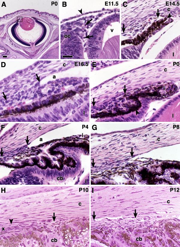
Figure 2.
Iridocorneal angle E11.5 to P12 Images from paraffin (A -G) and plastic (G-H) embedded B6 eyes of the indicated ages. (A) The box indicates the iridocorneal angle region that is illustrated at high power in the other panels of Figures 1 and 2. (B) At E11.5, loose mesenchymal tissue is present between the anterior edge of the optic cup (oc), the lens vesicle (v), and the surface ectoderm (arrowhead). Primitive vascular channels contain nucleated red blood cells (arrows). (C) At E14.5, two layers of epithelium form the OC region that will develop into the iris and ciliary body. The anterior layer is heavily pigmented (arrowhead). The arrows indicate the anterior and posterior extent of undifferentiated angle mesenchyme. The cornea (c) and lens (l) are well defined. (D) At E16.5, a small angle recess is present (a). The location of the future TM is evident (arrows). (E) In a newborn mouse, the mesenchyme of the developing iris (i) and TM (arrows) regions are distinguishable. The TM cells have elongated, more densely-staining nuclei and are arranged in lamellae (arrows). The ciliary processes (arrowheads) have begun to form. The angle recess is artifactually compressed in this image. (F) At P4, there is a long angle recess (a), and the iris and ciliary body (cb) are well formed. The cells of the future TM (arrows) show a dense lamellar arrangement. (G) At P8, the developing TM is less compressed than at earlier ages (arrows). (H) At P10, an endothelial lined vascular channel (arrowhead) is present at some locations. Intertrabecular spaces have begun to open in the anterior portion of the TM (arrow). The posterior aspect of the TM remains compressed (x). (I) At P12, A well-formed SC (arrows) is easily identified exterior to the posterior TM. Internal to SC, both anterior and posterior meshwork has become more open. Bars 200 μm (A) and 40 μm (B-I).

