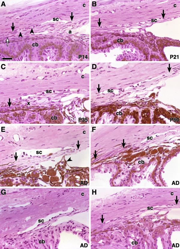
Figure 3.
Iridocorneal angle P14 to P63 Hematoxylin and eosin stained plastic sections from mice of the indicated ages. (A-D) strain B6. ( A) At P14, SC (arrows) contains vacuolar structures (arrowheads) that were confirmed to be giant vacuoles by EM (see below). The developing ciliary muscle is characterized by eosinophilic cytoplasm (open arrow). Intertrabecular spaces are obvious in the anterior TM and the deep angle recess (a) is present as a space between the anterior TM and iris root. c = cornea, cb = ciliary body. ( B) By P21, SC (arrows) extends from the posterior ciliary body to the end of Descemet's membrane. There are large spaces in the anterior TM. (C) By P35 there is further opening of the intertrabecular spaces that extend more posteriorly. The posterior TM (x) remains closely attached to the ciliary body, as it does in the adult. The ciliary muscle (arrow) consists of a few muscle fibers. (D) This P60 eye has a well developed SC (arrows) and TM and is very similar to that shown for P35. Comparison to older mice (up to 1 year old, not shown) indicates that the iridocorneal angle has reached maturity. The adult structure is similar in other mouse strains (E-H). All of the adult mice were approximately 63 days old. (E) A 129/SvEvTac mouse has a robust TM (arrows) and a broad SC. An iris process attaches to the anterior TM (arrowhead). (F) In this 129BS mouse, there is a robust TM and SC. The ciliary muscle (arrows) is particularly prominent in this strain. (G) BALB/cByJ. (H) In this DBA/2J mouse, SC is present but shows mild artifactual compression (arrows). Bar 40 μm.

