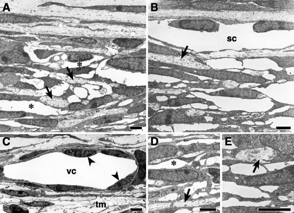
Figure 4.
Ultrastructure of the TM and SC in B6 mice at P10 (A, D) In the posterior TM, spaces are developing between the trabecular beams (asterisks). Small amounts of collagen and elastic tissue are demonstrated within the beams (arrows). Schlemm's canal is absent in these sections. (B, E) In the anterior TM, the spaces between adjacent trabecular beams are generally larger than posteriorly (compare B and E that are anterior to A and D that are posterior). SC is present in B but giant vacuoles are absent. Elastic tissue and collagen (arrows) are present in amounts similar to that of the posterior meshwork. (C) In a different level of section to B, SC is represented by a vascular channel (vc) adjacent to the differentiating TM (tm). The endothelial cells (arrowheads) lining this channel are less attenuated than in the adult SC and giant vacuoles are absent. Bars 1 μm.

