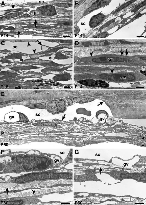
Figure 5.
Ultrastructure of the TM and SC in B6 mice from P14 to P60 ( A, B, D) are from the same P14 mouse. (A) At P14 the spaces in the posterior TM are smaller than at older ages (compare to a P18 eye in C). Trabecular beam collagen and elastic tissue (arrows) is more abundant than at younger ages. A Schwann cell (s) and accompanying myelinated nerve (arrowhead) are present close to SC. (B) In the anterior TM, there is a well-developed SC, but giant vacuoles are not common. There are fewer trabecular beams and larger intertrabecular spaces than in the posterior TM (compare to A, and see F, G). (C) The spaces (asterisks) between trabecular beams in this region of the posterior TM are more extensive than at P14. SC is lined by a thin endothelium (arrowheads) and contains giant vacuoles (arrow). (D) Smooth muscle cells (sm) lie internal to Schlemm's canal near its posterior termination. They are characterized by pinocytotic vesicles near the cell membrane (arrowheads), focal density of the plasma membrane (arrows) and cytoplasmic filaments (not seen at this magnification). (E) At P60, SC is lined by attenuated endothelium (arrows) and contains giant vacuoles (gv). a = anterior, p = posterior. (F) Partial segment of the posterior TM at P60. The trabecular beam extracellular matrix is dense. Collagen (arrow) is abundant while elastic tissue (arrowhead) is relatively sparse. (G) In the anterior TM, the beams are more delicate, and contain less extracellular matrix (arrow). A portion of the anterior iris (i) is present in this image. Bars 1 μm.

