Figure 6.
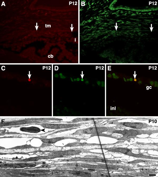
Absence of cell death in the developing iridocorneal angle. A double labeling assay that identifies fragmented DNA using fluorescently labeled dUTP (A, C) and detects chromatin condensation by binding of the dye YOYO-1 (B, D) was used to detect programmed cell death (PCD). Both assays were negative in a P12, B6 iridocorneal angle (A, B). The same was true for many sections at ages that spanned angle morphogenesis. i= iris, cb = ciliary body, arrows indicate the extremities of the TM (tm). (C, D, E) A cell undergoing PCD (arrow) is identified by double labeling in the retinal ganglion cell layer (gc) of the same eye shown in A and B. inl = inner nuclear layer. Dying retinal ganglion cells (RGCs) acted as internal positive controls for the PCD assays. Testis sections served as additional positive controls with each batch of processed slides, and abundant apoptotic cells were always detected. (F) Morphologic features of cell death were absent in the TM of a P10, B6 mouse. The trabecular cells demonstrate normal nuclei and normal cytoplasmic morphology. The same was true in many sections of eyes of different ages and strains. The iris (i) is resting against the inner edge of this central portion of the TM. A small lymphocyte (arrowhead) lies in the space between two trabecular beams. Bar 1 μm.

