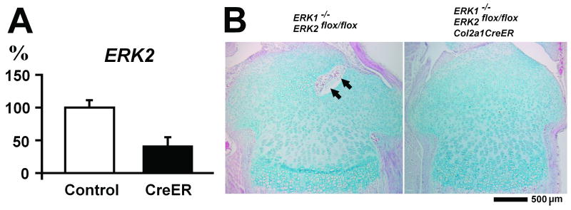
Figure 3.
ERK2 expression and histology of tibial epiphysis of ERK1-/-; ERK2flox/flox and ERK1-/-; ERK2flox/flox; Col2a1-CreER mice at P8 following tamoxifen injection at P4 and P6. A. Real time PCR analysis of ERK2 expression in the tibial epiphysis. Control: ERK1-/-; ERK2flox/flox (n=5), CreER: ERK1-/-; ERK2flox/flox; Col2a1-CreER (n=6). Values are the mean +/- SD. B. Hematoxylin, eosin, and alcian blue staining of the proximal tibia at P8. Arrows indicate vascular invasion in the developing secondary ossification center.

