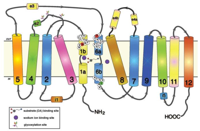
Fig. 1.
Two-dimensional representation of dopamine transporter topology based on LeuT structure. Twelve transmembrane domains are shown with helically unwound regions in the first and sixth domain; extracellular and intracellular loops include helical portions (e2, e3, e4a, e4b, and i1, 15, respectively) (see [122]). The residues mutation of which are discussed in the text are shown in colored circles: W84 in TM1b, red; D313 in TM6a, red; and Y335 in TM6b, green. The respective conformationally biased mutants are W84L and D313N (outward facing, [123]), and Y335A (inward facing, [126]).

