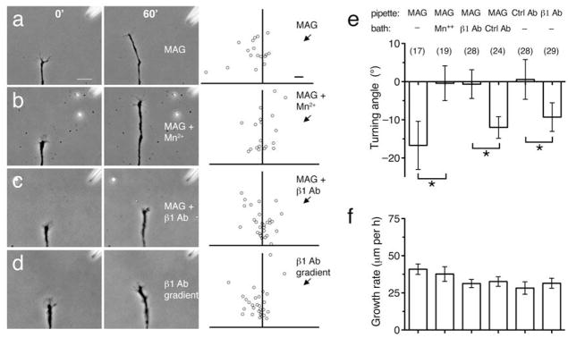
Figure 4. Polarized β1-integrin function mediates growth cone repulsion by MAG.
(a–c) Phase images show a representative growth cone at the onset (left) and the end (right) of a 1-hr exposure to a MAG gradient (150 μg/ml in the pipette). Assays were done in normal saline (a), saline containing Mn2+ (1 mM; b), or saline containing a function-blocking antibody to β1-integrin (5 μg/ml; c). Scatter plots depict the end points of individual growth cone extension for all the neurons examined for each condition. The origin is the center of the growth cone at the onset of the experiment and the original direction of growth was vertical. The arrow indicates the direction of the gradient. (d) Experiment carried out in the same manner as in (a), except that a gradient of function blocking antibody to β1-integrin was applied to the growth cone (0.4 mg/ml in the pipette). Scale bars, 20 μm (phase images) and 10 μm (scatter plots). (e,f) Summary of turning angles (e) and growth rates (f) for all conditions. Data are the mean ± s.e.m. (n = the number associated with each bar; * P < 0.05, bracketed comparisons, Mann-Whitney U-test).

