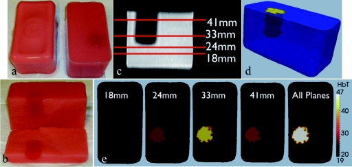
Figure 4.
Phantoms used (a) and (b) in experiment showing contrast recovery. Axial MR image of inclusion phantom (c) with locations of 4 imaging planes shown and text reporting imaging plane distance from bottom of phantom. 3D representation (d) of reconstruction using all 4 data sets simultaneously, and slices (e) through the inclusion after being reconstructed by individual data sets.

