
Fig. 7.
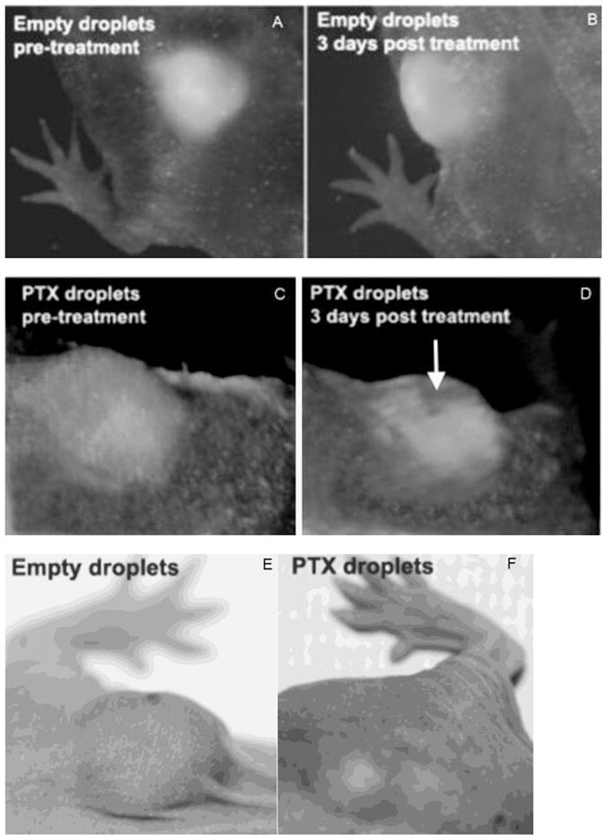
Intravital fluorescence images of subcutaneous pancreatic tumors before and after FUS treatment (A – D). Mice were injected with empty droplets (A, B) or PTX-loaded droplets (C, D). Fluorescence images of initial tumors are shown in panels A and C; images recorded three days after treatment are shown in panels B and D. Photographs of the tumors were taken 12 days after the treatment for empty droplets (E) and PTX-loaded droplets (F). Conditions of FUS treatment are presented in caption to Figure 6B. Mice were systemically injected with empty or PTX-loaded 1% PFCE/5%PEG-PDLA nanodroplets six hours before FUS treatment; DOX dose was 40 mg/kg.