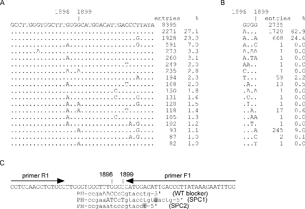
Figure 1. Target mutation site and assay design.

A non-contiguous megablast was performed using a query sequence of HBV nucleotide 1867–1926. A total of 8395 sequences were retrieved and the sequence patterns for the nucleotide 1881–1919 were compiled and sorted according to their number of entries and percentage representation (A). B, mutation patterns at nucleotide 1896–1899. C, assay design. Capital letters indicate LNAs. The 3’-end “PH” stands for phosphorylation. The nucleotide with fluorescence label was highlighted.
