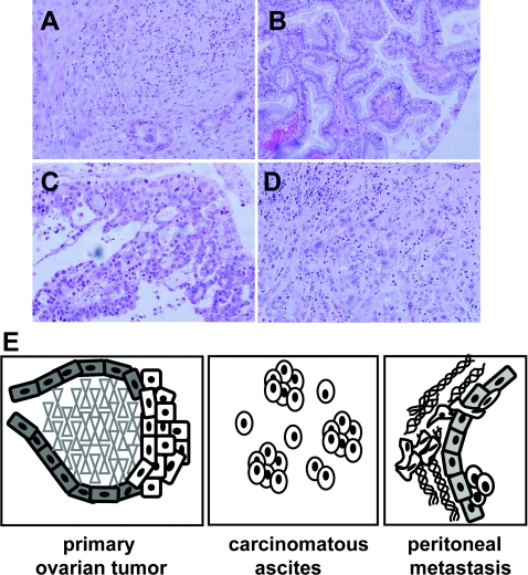
Figure 2. Ovarian tumours and the metastatic niche.
Examples of epithelial ovarian carcinoma histotypes after haematoxylin and eosin staining at ×200 magnification: serous (A), endometrioid (B), mucinous (C) and clear cell (D). (E) Model of ovarian cancer metastastatic niche. The model depicts a primary ovarian tumour arising from malignant transformation of ovarian surface epithelium. Single cells and MCAs are shed from the primary tumour into the peritoneal cavity. Accumulation of carcinomatous ascites is commonly observed, particularly in women with advanced disease. Metastasis is the result of multiple intraperitoneal adhesive events, whereupon tumour cells attach to peritoneal mesothelium, disrupt mesothelial cell–cell contacts and migrate into the submesothelial matrix to anchor secondary lesions on the bowel, diaphragm, omentum and other sites.

