
Fig. 8.
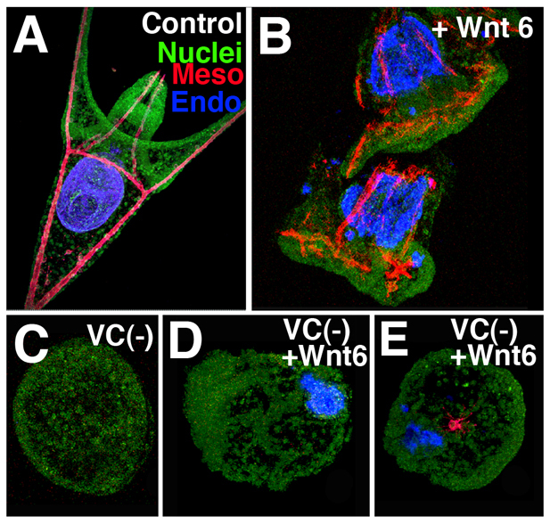
Wnt6 rescued endoderm in the VC(–) embryos. (A) Control pluteus larva viewed from below the anus. (B) Two embryos at the same stage as A overexpressing Wnt6. Each has enlarged and ectopic guts (blue) and mispatterned skeletal elements (red). (C) VC(–) control expressed no endomesoderm. (D) VC(–) embryos injected with Wnt6 mRNA expressed endoderm (n=32/32) or occasionally (E) endoderm plus one or two PMCs (n=3/32). SoxB1 staining (green) outlines the embryo.