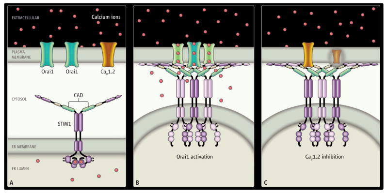
Figure. STIMulating.
STIM1 in the ER membrane activates Orai1 (green) and inhibits CaV1.2 (orange) in the plasma membrane. (A) The cell at rest. Calcium ions (red) are abundant in the extracellular space and within the ER lumen but are rare in the cytosol. Functional domains of STIM1 include the EF hand (with calcium ions bound), and the CRAC-activating domain (CAD, green). (B and C) ER-PM junctions when the cell is activated and Ca2+ is depleted from the ER lumen. Calcium ions unbinding from the EF hand of STIM1 trigger translocation of STIM1 to ER-PM junctions. The cytosolic junctional gap between ER and PM is sufficiently narrow (10 to 20 nm) to allow for direct molecular interaction of STIM1 with Orai1 and CaV1.2. The CAD of STIM1 recruits Orai1 to ER-PM junctions and opens Orai1 channels by direct binding to STIMulate Ca2+ influx (B). CaV1.2 channel proteins are also recruited to ER-PM junctions but are inhibited by the CAD interacting with the C terminus of CaV1.2 (C).

