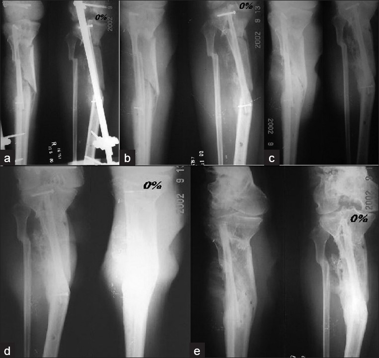
Figure 3.

(a) Postoperative X-rays (anteroposterior view) of a 22 year old male showing proximal tibial hemicortical defect. (b) Three months – 0% hypertrophy. (c) Six months later – 0% hypertrophy – note callus formation from residual periosteum. (d) Twelve months later – 0% hypertrophy – note callus formation from residual periosteum. (e) Twenty-four months later – 0% hypertrophy – note solid bridging callus
