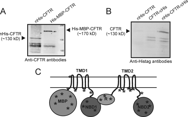
Fig. 1.
Expression of human CFTR in L. lactis. A, Membranes of L. lactis expressing His-MBP-CFTR and nHis-CFTR were analyzed by SDS-PAGE/Western blotting. CFTR was detected using anti-CFTR antibodies (clone 24–1, R&D systems). Ten micrograms protein was loaded per lane. B, Membranes of L. lactis expressing nHis-CFTR, CFTRcHis and nHis-CFTR-cHis were analyzed as described above, but now using anti-His-tag antibodies. C, Topology model of His-MBP-CFTR indicating the different domains, and showing positions of the tryptic peptides identified with LC-MS/MS. Peptides derived from all soluble domains of His-MBP-CFTR were found. For a complete list see supplemental Table S2 .

