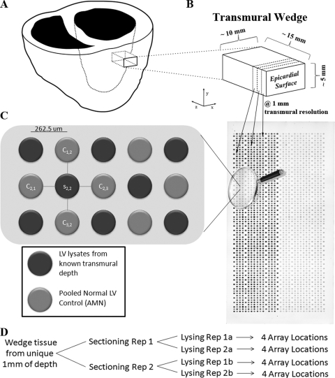
Fig. 1.
Experimental overview. A, A depiction of the acquisition of an anterior transmural wedge from a left ventricle (z axis represents the transmural dimension), which is immediately snap frozen. B, An individual transmural wedge being sectioned at 1-mm resolution (dotted line represents the sectioning plane x-y), at −24°C in a cryostat. Sample tissue from each disparate 1-mm region is lysed to create a whole-cell lysate. All 1-mm whole-cell lysates are then robotically printed on a RPMA in unique spots, as demonstrated by the arrows. C, A sypro ruby total protein stain of a single HRSH array is shown (the left side of the array contains samples printed at 600 μg/ml, and the right side at 150 μg/ml). The array is magnified to display the AMN format of RPMA printing. D, The structure of replicates printed on the RPMAs for each 1 mm transmural depth of wedge tissue. In total, sixteen replicates are printed that consist of independent sectioning and lysing replicates (reps) as well as repeated spotting on the array.

