Figure 3.
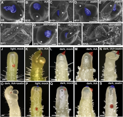
Induction of primordium formation and meristem tip growth by auxin and cytokinin. (A–I) Microapplication of auxin and cytokinin to dark-cultured apices. Dissected tomato apices were precultured in the light and transferred to darkness for 6 d. Lanolin containing 1% DMSO (A), 10 mM IAA (B,C), 1 mM zeatin (D–F), or 10 mM IAA plus 1 mM zeatin (G–I) was applied in the dark. These apices were further cultured in the dark for 10 d. (White asterisks) Pre-existing primordia. (F,I) Note that apices treated with 10 mM IAA plus 1 mM zeatin and with 1 mM zeatin alone continued to grow in the dark. (A,B) However, apices treated with 1% DMSO or 10 mM IAA did not grow. (C,E,H) In addition, 10 mM IAA alone, 10 mM IAA plus 1 mM zeatin, and 1 mM zeatin alone promoted the development of pre-existing P1 and I1. The numbers in the bottom left corner show the number of apices that show the displayed phenotype out of the total number of samples. Thus cytokinin induces leaf initiation in the dark, and auxin promotes leaf initiation in the presence of cytokinin. (J–O) Microapplication of auxin and cytokinin to the flank of the meristems of tomato NPA pins. Dissected apices were cultured in the presence of NPA. Resulting pin-shaped apices (NPA pins) were precultured in the light or the dark, and microapplication of IAA and cytokinin was performed. Microapplication of 1% DMSO (mock) lanolin in the light (J), 10 mM IAA lanolin in the light (K), 1% DMSO lanolin in the dark (L), 10 mM IAA lanolin in the dark (M), 10 mM IAA plus 1 mM zeatin lanolin in the dark (N), and 10 mM IAA plus 10 mM zeatin lanolin in the dark (O). (P–S) Microapplication to the flank and the summit of the meristem of NPA pins. Microapplication of 1% DMSO lanolin to the flank and the summit in the light (P), 1% DMSO lanolin to the flank and the summit in the dark (Q), 10 mM IAA lanolin to the flank and 1% DMSO lanolin to the summit in the dark (R), and 1 mM zeatin lanolin to the summit and 1% DMSO lanolin to the flank in the dark (S). (A–I) Scanning electron microscope images. (J–S) Stereomicroscope images. Lanolin dots applied to the flank are colored red, and those applied to the summit are colored blue. Bars, 100 μm.

