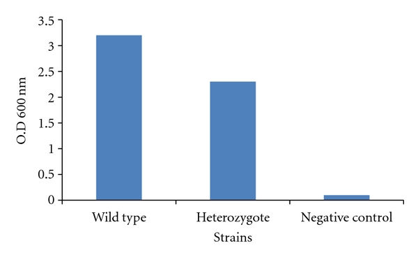
Figure 3.

Biofilm formation on polystyrene surfaces. Strains were plated on polystyrene microtiter plates and incubated for 24 h followed by rinsing and methanol addition to allow the fixation of biofilm-forming cells. Afterward, crystal violet was added to stain the newly formed biofilm cells. Unbound crystal violet was washed away and bound crystal violet was released by adding acetic acid to the wells. OD at 600 nm of the released crystal violet was read, and it proportionally corresponded to the amount of biofilm forming cells present in the wells. The negative control lacked any C. albicans cells but was treated similarly. Note the significant defect in biofilm formation in the mutant strain (P = .002).
