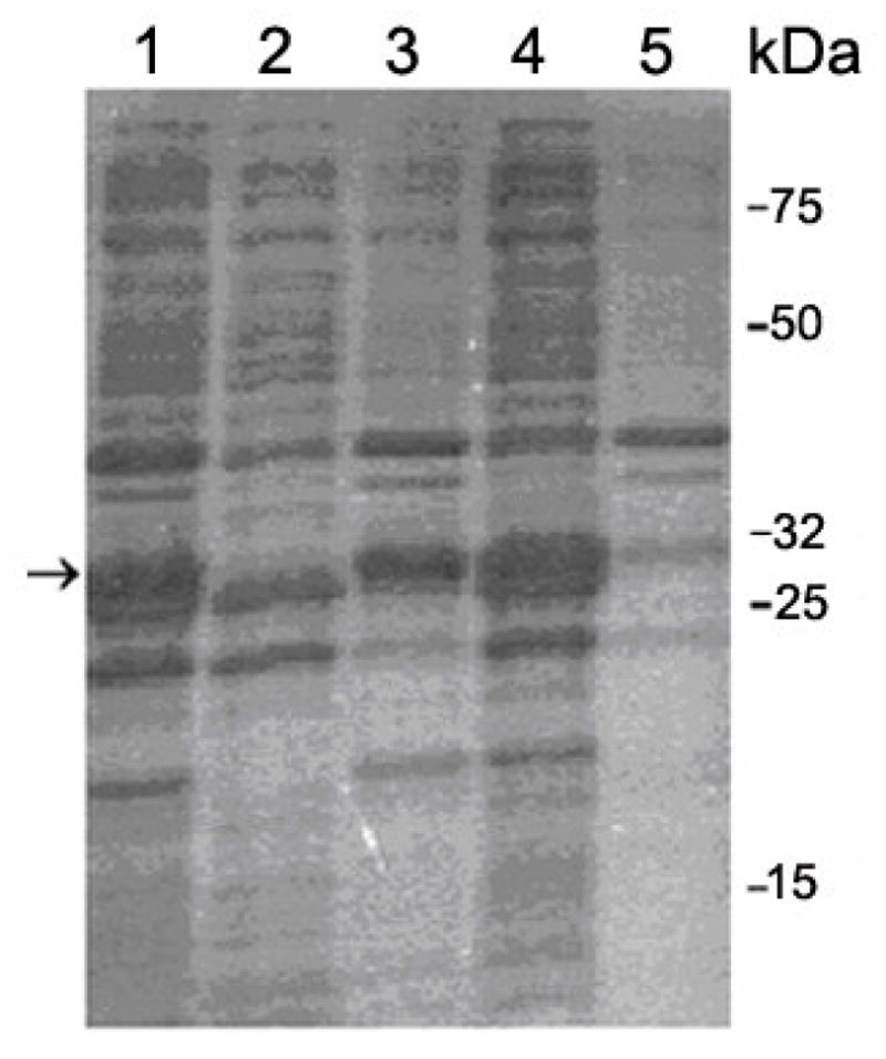
Figure 2.

15% SDS-PAGE analysis of the cells extracts. Lane 1: total lysate treated with 1% (w/v) SDS. Lane 2: soluble supernatant from cells treated with 1% Triton X-100 (v/v). Lane 3: insoluble pellet from cells treated with 1% Triton X-100 (v/v). Lane 4: soluble supernatant from cells treated with 0.5% sodium lauroyl sarcosinate and 0.8% Triton X-100 (v/v). Lane 5: insoluble pellet from cells treated with 0.5% sodium lauroyl sarcosinate and 0.8% Triton X-100 (v/v). It is clear from the data in Lane 4 and Lane 5 that most of the fusion protein was successfully solubilized. The arrow indicates the position of GST-Aβ having the molecular mass of ~ 31 kD
