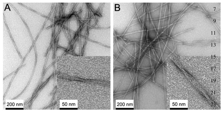
Figure 6.
Negatively stained transmission electron micrograph (TEM) images of Aβ(1–40) fibrils after 1 week of the incubation at room temperature with agitation for (A) recombinant Aβ(1–40) and (B) synthetic Aβ(1–40). The insets show magnified images. The observed morphologies and the width are similar between synthetic and recombinant Aβ peptides.

