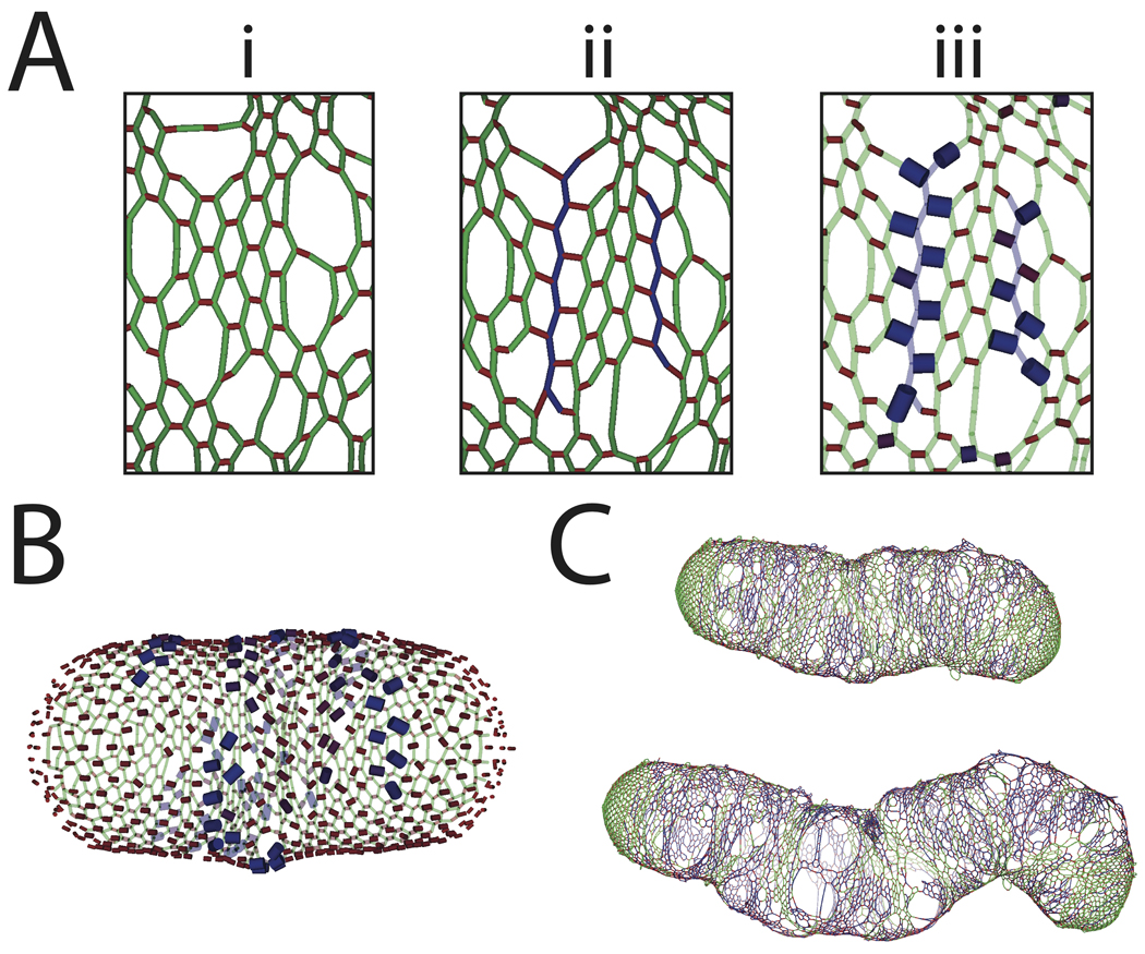
Figure 7. Tension-sensitive insertion causes rapid loss of cell shape.
(A) Schematic of likelihood of initiation surrounding a recently inserted glycan strand based on tension-sensitive insertion. (i) Initial network before insertion, (ii) relaxed network after the insertion of two new strands (blue), (iii) peptides with largest tension highlighted by increased width and blue shading. (B) Schematic of likelihood of initiation across the cell wall, with increased insertion probability at crosslinks with highest tension. (C) Cell wall grown to two- and three-fold its original mass, with significant bending and increases in local width. After three-fold growth, the cell wall has almost completely lost its rod shape.

