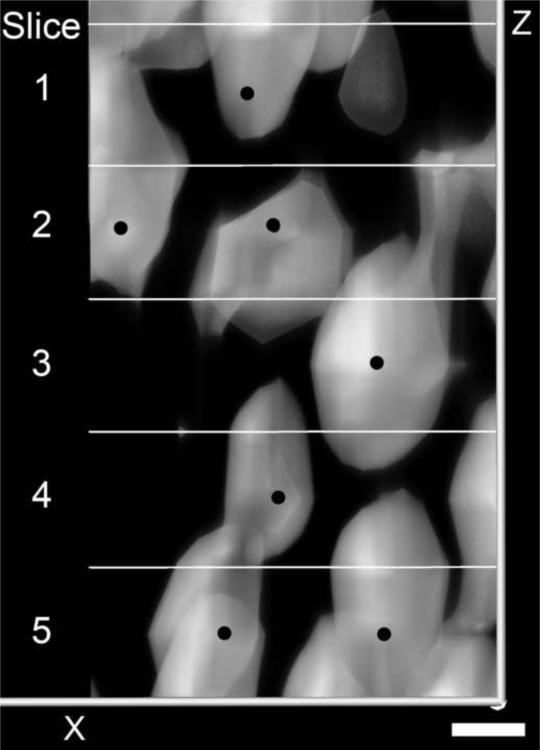
Fig. 2.
This is direct volume rendering of a portion of the RC containing at least seven SGNs (black dot) that were counted in five, 10 μm sections (panels labeled 1–5). Notice that although the cells spanned across different optical sections they could be marked and identified to insure that they were only counted once. Bar indicates 5 μm.

