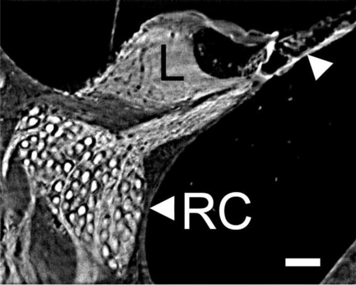
Fig. 4.
This is a higher magnification view (scala media cross section) of a midmodiolar cross section through the whole cochlea imaged by TSLIM. Note that image can be further enlarged within Amira and on a computer screen to view details of the cells and tissues of the scala media. Even at this magnification one can easily resolve Rosenthal's canal (RC) containing SGNs, the spiral limbus (L), and the outer hair cells (arrowhead). Bar indicates 50 μm.

