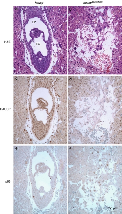
Figure 2.
Analysis of embryos of day E7.5 from hauspΔEx6/+ intercross. Two representative embryos are shown: an HAUSP staining positive embryo (hausp+) in a, c, and e; and an HAUSP staining negative embryo (hauspΔEx6/ΔEx6) in b, d, and f. The hausp+ embryo showed formation of germinal layers and characteristic structures of exocoelom (EC) and ectoplacental cavity (EP) for normal embryos at day E7.5 (a), positive staining for HAUSP (c), but little or no p53 staining (e), whereas the hauspΔEx6/ΔEx6 embryo showed no recognizable structures (b), no HAUSP staining in the remaining embryonic cells (d), and detectable p53 staining in all remaining embryonic cells (f)

