Figure 5.
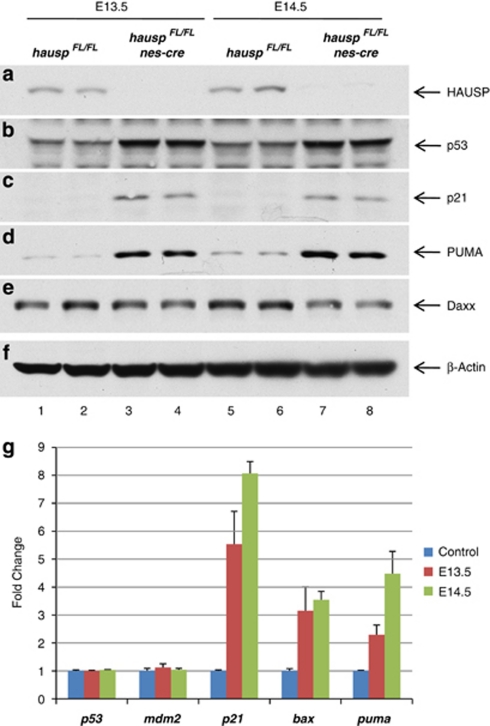
Depletion of HAUSP in neural cells led to accumulation of p53 and activation of p53 target genes. Protein extracts were prepared from cortex of the brains collected at days E13.5 and E14.5. The protein levels of HAUSP (a), p53 (b), p53 downstream target p21 (c), PUMA (d), Daxx (e), and total proteins represented by β-actin (f) were determined by western blot. Protein extracts from two individual hauspFL/FL brains at E13.5 (lanes 1 and 2) and at E14.5 (lanes 5 and 6), along with protein extracts from two individual hauspFL/FL; nes-cre brains at E13.5 (lanes 3 and 4) and at E14.5 (lanes 7 and 8), were analyzed. HAUSP protein was shown to be nearly depleted in hauspFL/FL; nes-cre brains (a; lanes 3, 4, 7, and 8) compared with HAUSP protein in the control brains (a; lanes 1, 2, 5, and 6). In contrast, the protein level of p53 was elevated in hauspFL/FL; nes-cre brains (b; lanes 3, 4, 7, and 8 versus lanes 1, 2, 5, and 6) and so were the p53 downstream targets, p21 (c) and PUMA (d). Daxx, a reported HAUSP substrate, was slightly reduced after knockout of hausp (e; lanes 3, 4, 7, and 8 versus lanes 1, 2, 5, and 6). Total proteins were comparable, indicated by the protein levels of β-actin in each lane (f). (g) Quantitative PCR analysis of transcriptional levels of p53, mdm2, p21, bax, and puma in hauspFL/FL; nes-cre brains collected at days E13.5 and E14.5. The hauspFL/+; nes-cre brains were used as controls. Whereas little or no change of transcription was observed for p53 and mdm2, the transcription levels of p53 target genes, p21, bax, and puma were significantly increased in hauspFL/FL; nes-cre brains. The error bars represent S.D.

