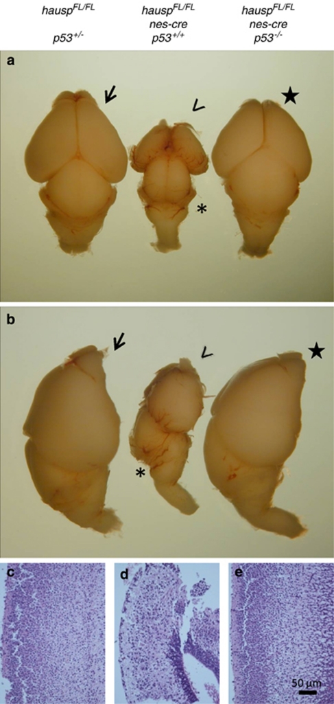
Figure 7.
Deletion of p53 in hauspFL/FL; nes-cre mice restored the cell density in the cortex and the size of the brain. Dorsal (a) and lateral (b) views of the brains collected at day E18.5 from hauspFL/FL; p53+/− (arrow), hauspFL/FL; nes-cre (arrowhead), and hauspFL/FL; p53−/−; nes-cre (star) are shown. The hauspFL/FL; nes-cre brain (arrow head) was much smaller than the control brain (arrow), displaying little or no development of cerebellum (asterisk), whereas the hauspFL/FL; p53−/−; nes-cre brain (star) showed comparable size to that of the control brain (arrow), with similar development of cerebellum. (c–e) The sagittal sections of the brains were stained with hematoxylin and eosin. The thickness and cell density of the cortex of the hauspFL/FL; p53−/−; nes-cre brain (e) was comparable to that of the control hauspFL/FL; p53+/− brain (c), whereas the hauspFL/FL; nes-cre brain (d) showed much reduced thickness and much lower cell density than that of the control brain (c). These results suggested that concomitant deletion of p53 partially rescued the developmental defects in hauspFL/FL; nes-cre brains

