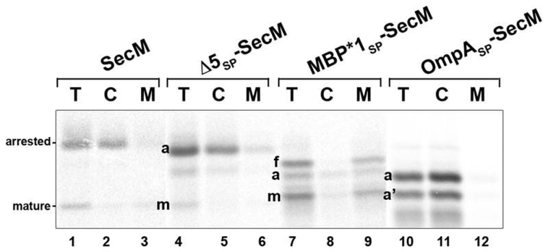
Fig. 2.

Localization of translation-arrested SecM RNCs. MNY3 transformed with a plasmid that encodes the indicated SecM derivative were pulse labeled. Cells were converted to spheroplasts, which were then separated into cytoplasmic and membrane fractions, and full-length (f), arrested (a) and mature (m) forms of SecM were immunoprecipitated from each fraction. A SecM fragment that was presumably a nascent chain attached to the ribosome situated behind the stalled ribosome (a′) was also immunoprecipitated from some of the samples. T, total spheroplast extract; C, cytoplasm; M, membranes.
