
Figure 2.
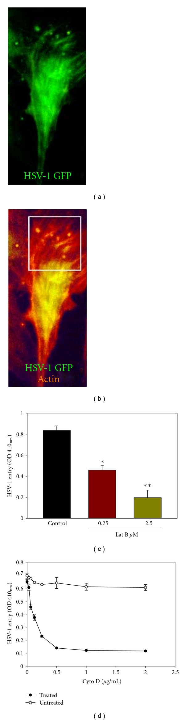
HSV-1 entry into hMSCs is cytoskeleton dependent. (a) An infected cell showing the virus particles (green). Cells were infected with HSV-1 (K26GFP) at 50 PFU/cell and imaged 45 min postinfection. (b) Virus particles colocalize with F-actin in filopodia-like structures (boxed area). F-actin was stained by rhodamine phalloidin (red). Areas where virus particle (green) and F-actin overlap appear yellow. (c) HSV-1(KOS) gL86 entry into hMSCs (50 PFU/cell) was inhibited by actin depolymerizing agent Latrunculin B (Lat B) in a dose-dependent manner. (d) HSV-1(KOS) gL86 entry into hMSCs (50 PFU/cell) was inhibited by actin depolymerizing agent Cytochalisin D (Cyto D) in a dose-dependent manner. The mock-treated hMSCs were used as a control. Viral entry was quantitated 6 h after infection at 410 nm using a spectrophotometer *P < .05, **P < .01.