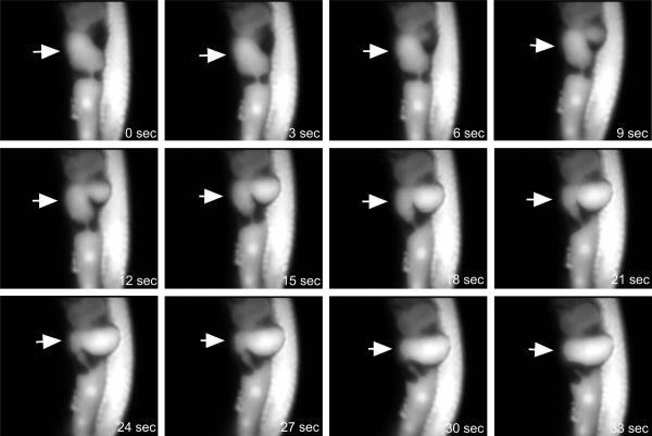
Figure 3.
Fluorescence of Calcium Green-1, dextran 10,000 MW in a fertilized oocyte monitored by CCD camera. Images are presented at 3 sec intervals. The oocyte that is fertilized is denoted by the arrow. In this image, the uterus is above the fertilized oocyte, and contains a 2-celled embryo. An immature oocyte is below the fertilized oocyte, and has not yet undergone nuclear envelope breakdown. At t = 6 sec, the mature oocyte begins to enter the spermatheca through the sphincter at its entrance (at this point the oocyte is not yet encased in eggshell and retains flexibility). Between t = 12 sec and t = 15 sec, the leading edge of the oocyte has engulfed a sperm leading to a local ~ 30% increase in fluorescence, i.e., cytosolic [Ca++]. As the oocyte enters the spermatheca, the [Ca++] elevation spreads throughout the cell. At t = 30 sec, the entire oocyte has entered the spermatheca, and the oocyte has become rigid as a result of the formation of eggshell.

