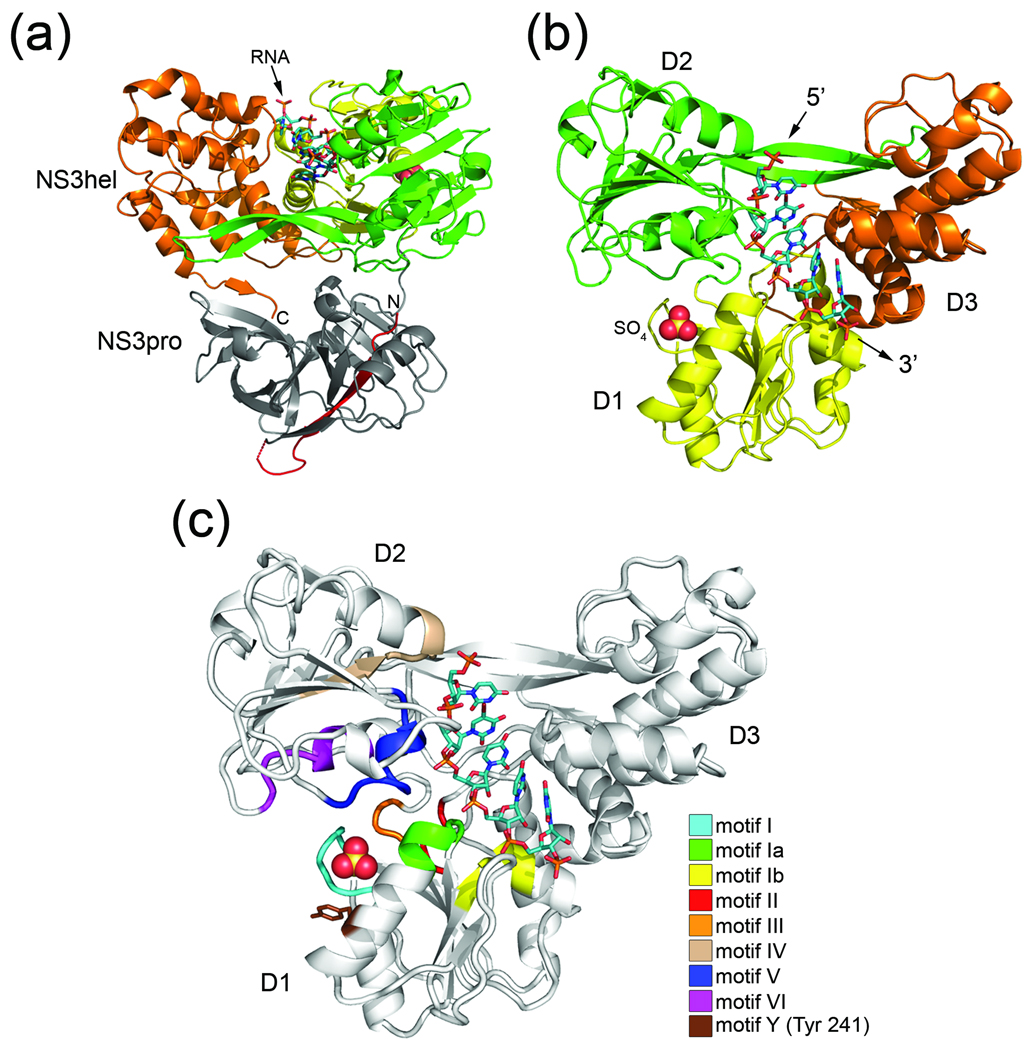
Fig. 1.
Structure of Full-length HCV NS3 bound to RNA. (a) Cartoon representation of NS3 viewed from the side. NS3hel domains 1 (D1), domain 2 (D2), and domain 3 (D3) are colored yellow, green, and orange, respectively. NS3pro (gray) with N-terminally-fused NS4A peptide (red) lies beneath NS3hel in this orientation. N and C termini and ssRNA are labeled accordingly. (b) A top view of NS3 (colored as above). NS3pro is removed for clarity. The bound sulfate ion is shown as a space-filling model, while the 5’ and 3’-ends of the ssRNA (stick model) are labeled accordingly. (c) Conserved SF2 helicase motifs mapped onto NS3. Motif I (residues 204–211), motif Ia (residues 230–235), motif Ib (residues 268–272), motif II (residues 290–293), motif III (residues 322–324), motif IV (residues 365–372), motif V (residues 411–419), and motif VI (460–467) are colored according to the legend. Tyr 241 (motif Y) is shown as a stick model.

