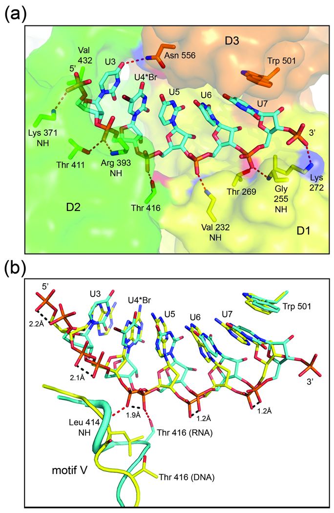
Fig. 3.
HCV NS3 binding to single-stranded nucleic acid. (a) Close-up view of ssRNA bound to NS3. RNA is shown as sticks colored by atom type (carbon, cyan; nitrogen, dark blue; oxygen, red; phosphate, orange; bromine, burgundy). NS3 is shown as a transparent surface and colored by domain (as in Fig. 1). The side chain or backbone atoms of NS3 residues interacting with the RNA are represented by sticks and are labeled accordingly. The side chains of Lys 371 and Arg 393 are disordered in the structure and omitted here for clarity. Nucleotide residues (U3–U7) and the 5’ and 3’-ends of the strand are labeled. (b) Overlay of the NS3-rU8Br4 and NS3hel-ssDNA (PDB ID: 3KQH) structures. RNA is shown as above, while the DNA is shown with carbons colored yellow. Protein residues in motif V of NS3-rU8Br4 (cyan) and NS3hel-ssDNA (yellow) are shown as cartoon coils. Leu 414 of NS3hel-ssDNA and the side chain atoms for Thr 416 and Trp 501 in both structures are represented by sticks. Hydrogen bonds between protein atoms and the phosphodiester backbone linking nucleotides 4 and 5 are represented by red dashed lines. Distances between corresponding phosphoryl oxygens of DNA and RNA are indicated by dashed lines (black).

