Figure 2.
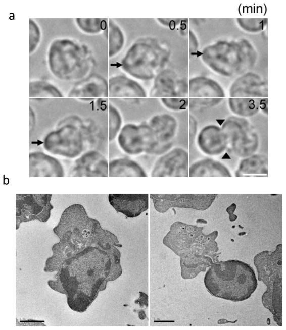
During enucleation the nucleus is rapidly squeezed out from the cell, forming a bleb-like structure. (a) Time-lapse live-cell imaging of late erythroblasts undergoing nuclear extrusion. The nucleus is quickly squeezed out from a limited area of the cortex facing the nucleus, forming the bleb-like structure (arrows). Note that the nucleus becomes largely deformed during enucleation, and that a cytokinetic-like furrow is observed at the neck region of the bleb-like protrusion (arrowheads). Scale bar, 5 μm.(b) Electron microscope images of late erythroblasts undergoing enucleation in an in vitro culture system. Shown are late erythroblasts at early (left) and late (right) stages of nuclear extrusion. These images show deformed nuclei and cytokinesis-like furrows similar to what has been seen in erythroid cells from mice [10], indicating that late erythroblasts in the in vitro culture system reproduce the in vivo situation. The images were taken by T. Ramirez and M. Murata-Hori. Scale bar, 2 μm.

