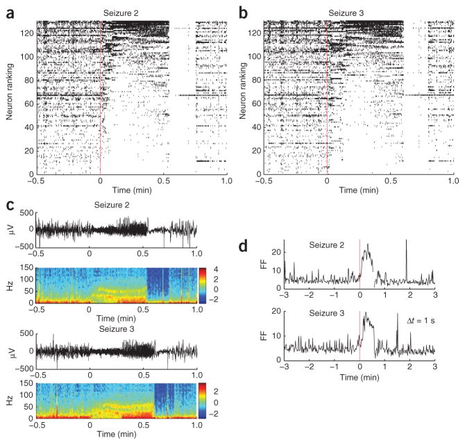
Figure 3.
Reproducibility of neuronal spiking modulation patterns across consecutive seizures. (a,b) An example from participant A with 131 neurons. Following conventions used in Figure 1c, neurons are ranked according to their mean rates measured during the seizure. Seizure 3 (b) follows the same ranking as seizure 2 (a); that is, the single units in any given row of seizures 2 and 3 are the same. Most neurons coarsely preserved the types of spiking rate modulation across the two seizures. For example, the lowest-ranked neurons decreased or stopped spiking; and many of the top-ranked neurons presented similar transient increases in spiking rate modulation. As in seizure 1 (Fig. 1), an almost complete suppression of spiking in the neuronal population occurred abruptly at seizure termination. (c) The corresponding low-pass filtered local field potentials (LFPs) and spectrograms (from the same microelectrode array channel shown in Fig. 1; power in dB). (d) The Fano factor for the spike counts (1-s time bins) in the population of recorded neurons showed similar increase during both seizures, reflecting the increased heterogeneity in neuronal spiking across the population.

