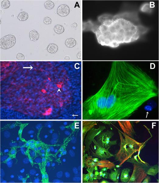
Fig. 1.
Confirmation of stem cell identity and multipotent differentiation capability of the cultured mouse embryonic stem cells (MESCs) in feeder-layer-free culture system. A: Phase-contrast image of cultured MESCs without a feeder layer maintaining their typical round colonies with smooth edges indicating lack of differentiation. B: Positive staining with stem cell specific marker SSEA-1. C: Positive staining for the neuronal cell marker: neurofilament (NF). Antibodies to NF stain both cell bodies (arrowhead) and axons (large arrow). Nuclei are stained blue with DAPI (small arrow). D: Positive staining for the cardiac cell marker: cardiac Troponin T (cTnT) (green stain). Organized myofibrils are seen. E: Positive staining for the skeletal muscle cell marker: fast skeletal Troponin T (fsTnT) (green stain). DAPI, a blue nuclear fluorescent dye shows nuclear staining. F: Positive staining for both a-actinin (green) and desmin (red), markers for mesoderm-derived cell types. Magnifications for Figure 1 taken are: (A) 100×; (B) 250×; (C) 100×; (D) 600×;(E) 250×;(F) 250×.

