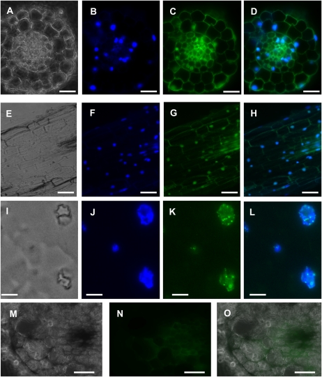
Fig. 4.
Ntann12 immunolocalization in tobacco root sections visualized by fluorescence microscopy. (A–D) A 100μm cross-section. (E–H) A 100 μm longitudinal section. (I–L) A 50–70 nm cross-section. (M–O) A 100μm cross-section. (A), (E), (I), (M) Phase contrast. (B), (F), (J) Staining with 4',6-diamidine 2-phenyl indole (DAPI). (C), (G), (K) Immunostaining with affinity-purified antiserum against Ntann12. (D), (H), (L) Superposition of DAPI staining and immunostaining with affinity-purified antiserum against Ntann12. The green fluorescence is indicative of the presence of Ntann12, whereas DNA is represented by blue colour resulting from DAPI staining. (N), (O) The green fluorescence shows the background autofluorescence without immunostaining. Bar=50 μm (A–H and M–O), 5 μm (I–L).

