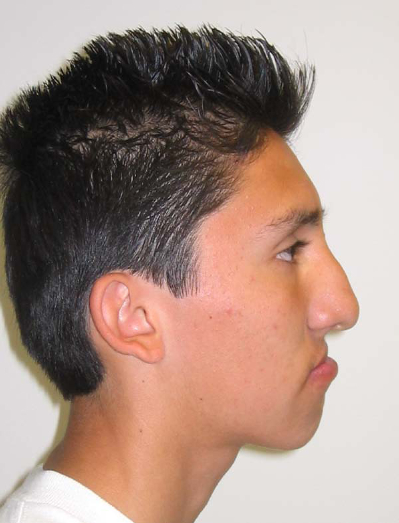
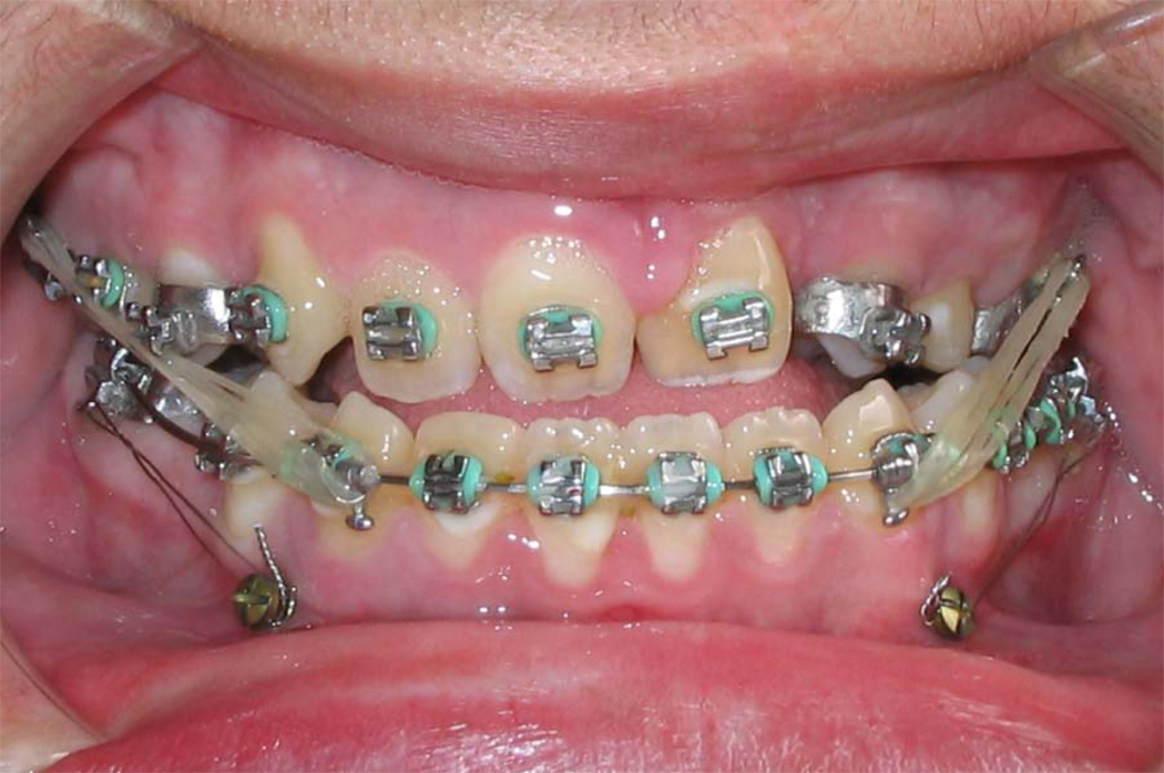
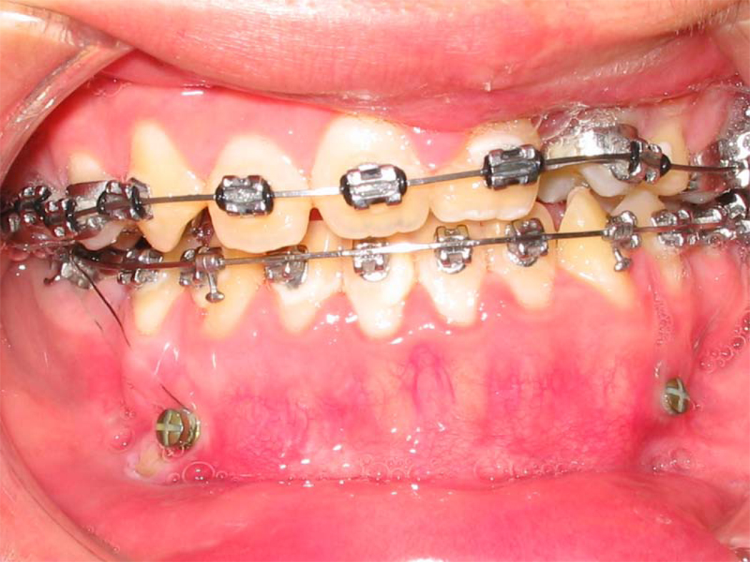
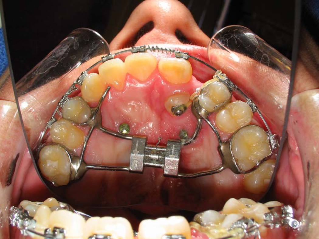
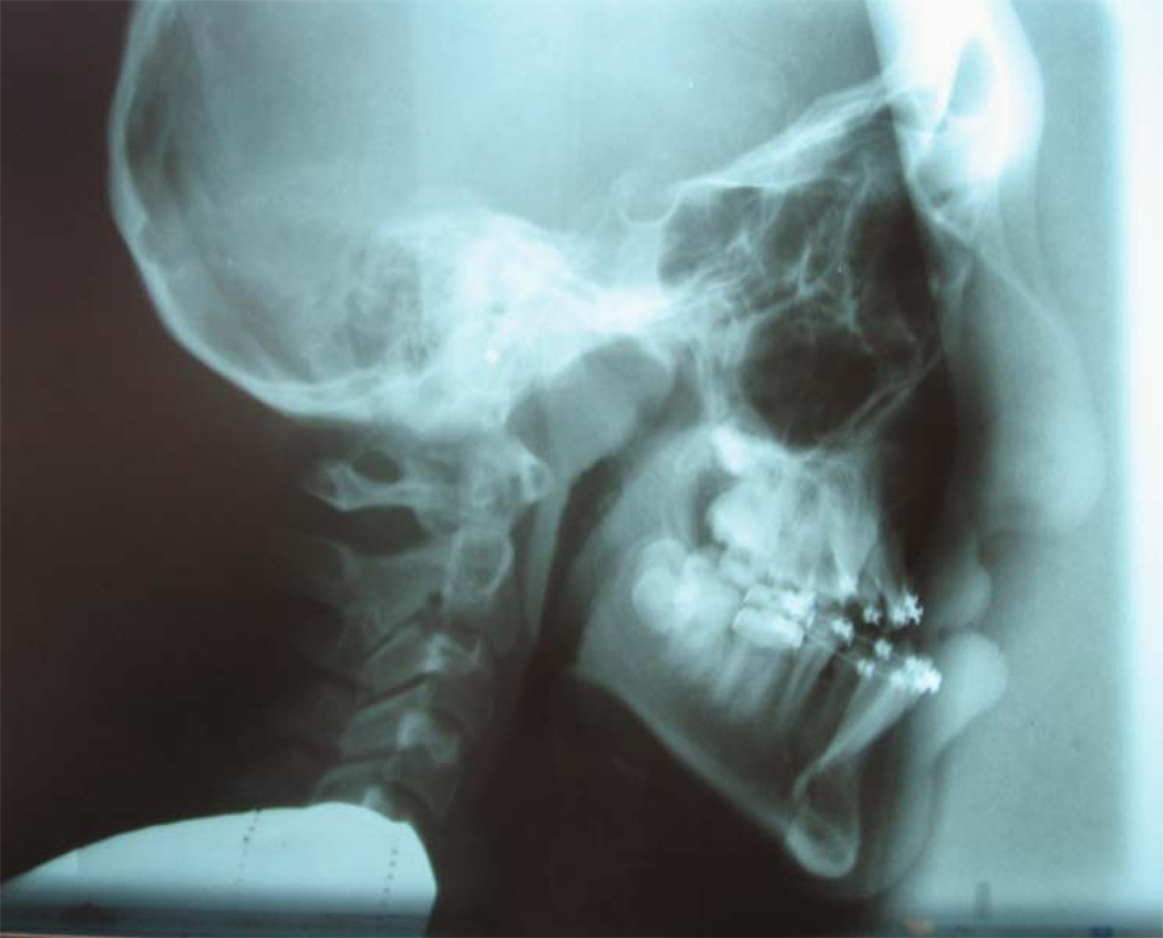
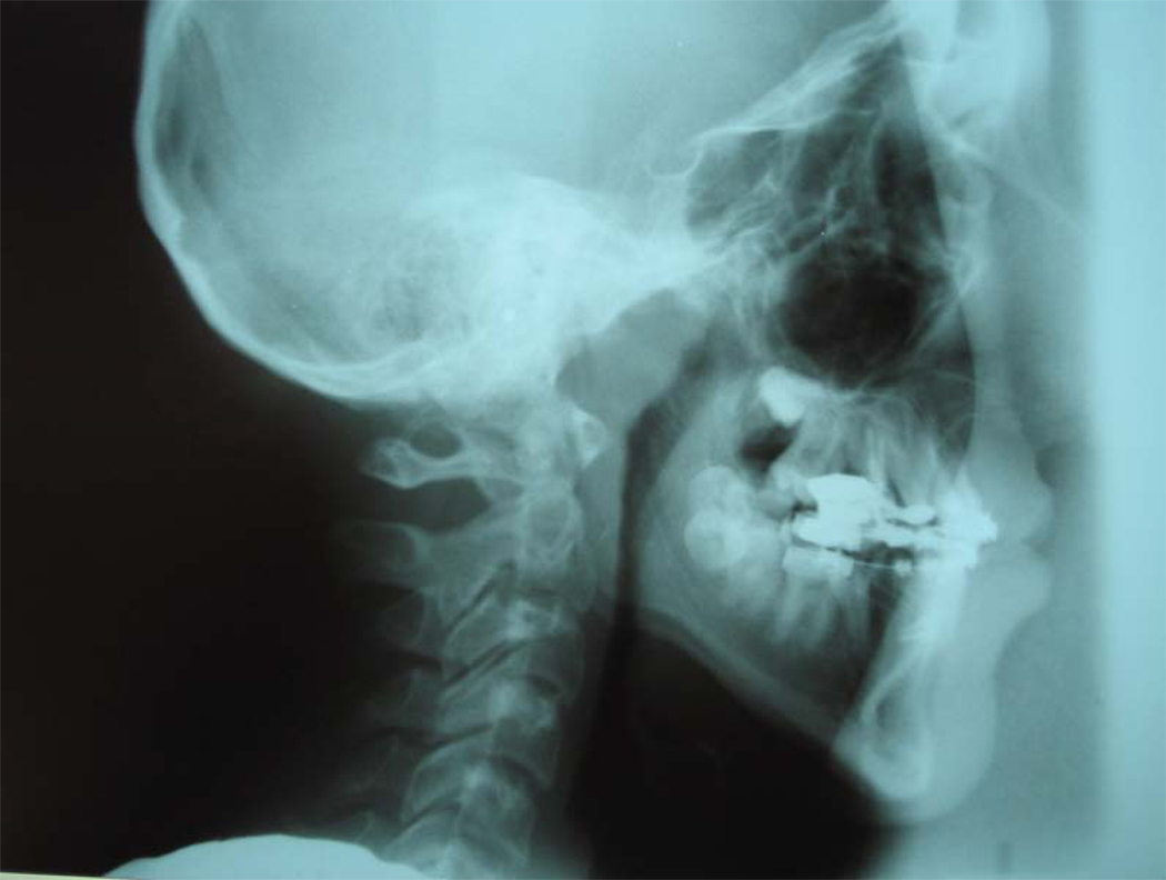
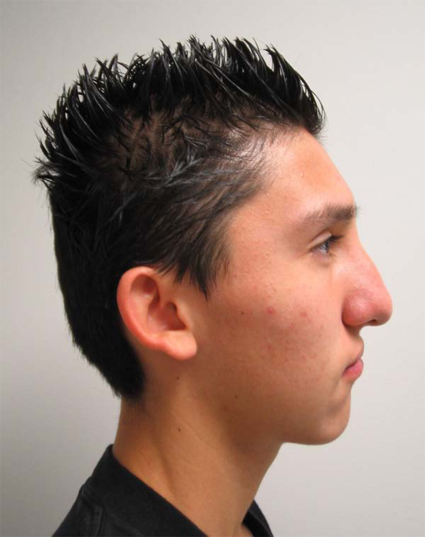
Figure 5.
Microimplant-supported maxillary protraction. (A) Extraoral profile view; (B) intraoral frontal view showing lower microimplant position; (C) intraoral frontal view showing treatment changes; (D) palatal occlusal view showing microimplant positions; (E) pre-treatment lateral cephalogram; (F) post-treatment cephalogram after four months of maxillary protraction; (G) post-treatment extraoral profile view. (Color version of figure is available online.)

