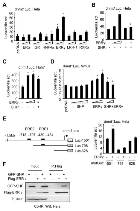
Fig. 1.
SHP inhibition of dnmt1 promoter activity by ERRγ. (A) Transient transfection assays to determine dnmt1 promoter luciferase (luc) activity (act) by expression of ERα (50, 200 ng), GR (50, 200 ng), HNF4α (50, 200 ng), ERRγ (50, 200 ng), LRH-1 (50, 200 ng), or RXRα (50, 200 ng) in Hela cells. (B–D) Transient transfection assays to determine dnmt1 promoter luciferase activity by co-expression of ERRγ and SHP in Hela (B) (ERRγ: 50, 200 ng; SHP, 200 ng; ERRγ+SHP, 200 ng+200 ng), Huh7 (C) (ERRγ: 100, 400 ng; ERRγ+SHP: 400 ng+100 ng), and Nmuli cells (D) (SHP: 100, 200, 400, 800 ng; ERRγ: 200, 400, 800 ng; ERRγ+SHP: 800 ng+200 ng, 800 ng+400 ng). (E) Left: Diagram showing the location of EREs in the dnmt1 promoter and its deletion constructs. Right: Transient transfection assays to determine dnmt1 promoter lucifease activity by ERRγ (100 ng) in Hela cells. A–E: The luciferase activities were normalized by renilla activities. Data is represented as mean ± SEM (*p<0.01 vs. control; ¥p<0.01 vs. ERRγ alone). The experiments were repeated three times (triplicate wells/time) with similar results. One representative result is shown. (F) Immunoprecipitation (IP) and Western blots (WB) to determine the direct association of SHP with the ERRγ protein. Hela cells were transfected with Flag-ERRγ (1 μg, 3 cm plate) and/or GFP-SHP (1 μg, 3 cm plate) expression vectors. Anti-Flag antibodies were used to immunoprecipitate ERRγ, and the protein levels of SHP and ERRγ were detected by WB using anti-GFP or anti-Flag antibodies, respectively.

