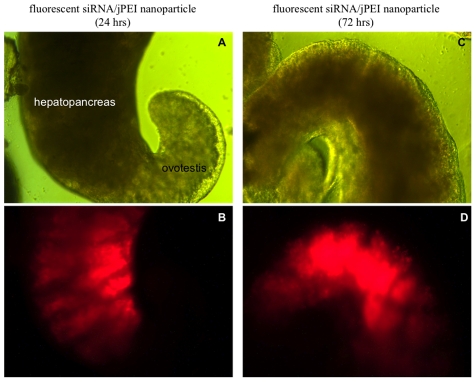
Figure 1. Snail organs transfected with the jPEI/fluorescent siRNA nanoparticles visualized by light and fluorescence microscopy.
Panels A and C: Images of hepatopancreas and ovotestis regions of juvenile snails that were soaked either in jPEI/Alexa 555 siRNA for either 24 or 72 hrs viewed without fluorescence (10× magnification). Panels B and D: Images of the hepatopancreas and ovotestis tissues shown in panels A and C, subjected to fluorescence microscopy as described in Materials and Methods. Note the intense flourescence (red stain) in the hepatopancreas compared to the ovotestis, indicating preferential uptake of the jPEI/fluorecent siRNA nanoparticles into this tissue.

