
Figure 2. EGF reduces NGAL expression and its effect via EGFR.
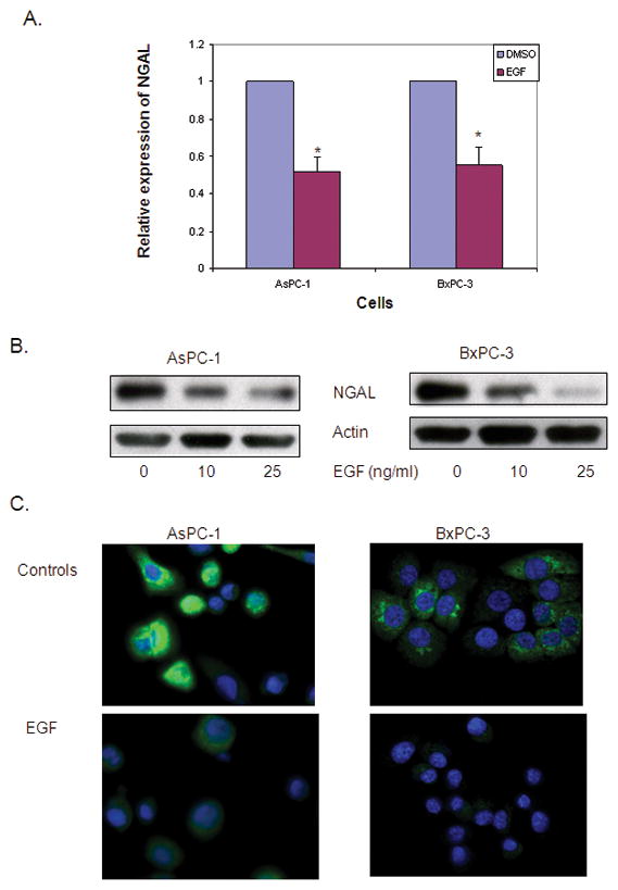
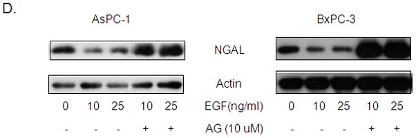
A. Quantitative real-time PCR for NGAL expression using total RNA extracted from cells that were treated with 25 ng/ml of EGF for 24 hours. The NGAL expression was determined using the formula of 2−ΔΔCT and expressed as means ± SE of three independent experiments. * represents p<0.05. B. Western blot analysis revealed NGAL expression in cells treated with the indicated concentration of EGF for 24 hours. C. Immunofluorescent staining of NGAL. Cells were treated with 25 ng/ml of EGF in serum free media. Twenty-four hours later, cells were stained for NGAL (green) and nuclei (DAPI, blue) and photographed using a confocal microscope at ×60 magnification. D. Cells were treated with or without AG1478 for 1 hour and followed by EGF treatment at the indicated dose for 24 hours. NGAL protein levels were determined by western blot analysis.