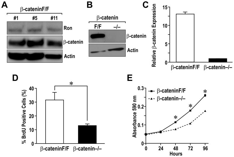
Figure 4. β-catenin deletion in breast cancer cell lines leads to a decrease in cell proliferation in vitro.
A, Ron and β-catenin expression was analyzed by Western in three independent mammary tumor cell lines (β-cateninF/F) derived from MMTV-Ron expressing mice containing a Floxed β-catenin allele. Actin expression was used as a loading control. B, The β-cateninF/F cells were infected with an adenovirus expressing Cre recombinase and GFP. GFP-positive (β-catenin−/−) and negative (β-cateninF/F) cells were isolated by FACS sorting and analyzed for β-catenin expression Western analysis. C, Quantative real-time PCR was utilized to examine β-catenin mRNA expression in β-cateninF/F and β-catenin−/− cells. Relative mRNA levels are shown normalized to an internal control. D, Deletion of β-catenin leads to decreased proliferation. β-cateninF/F and β-catenin−/− cells were incubated with BrdU for 4 hours and immunocytostaining for BrdU incorporation was performed. The percent of cells straining positive for BrdU was quantitated from three independent high power fields and the data tabulated is a result from three independent experiments. *, P < 0.05. E, β-cateninF/F and β-catenin−/− cells were plated in triplicate in 24 well plates. Cell number was measured at 0, 24, 48, 72 and 96 hours by crystal violet staining assays. The graph is representative of the three independent experiments. *, P < 0.05 compared to the corresponding β-catenin−/− cells.

