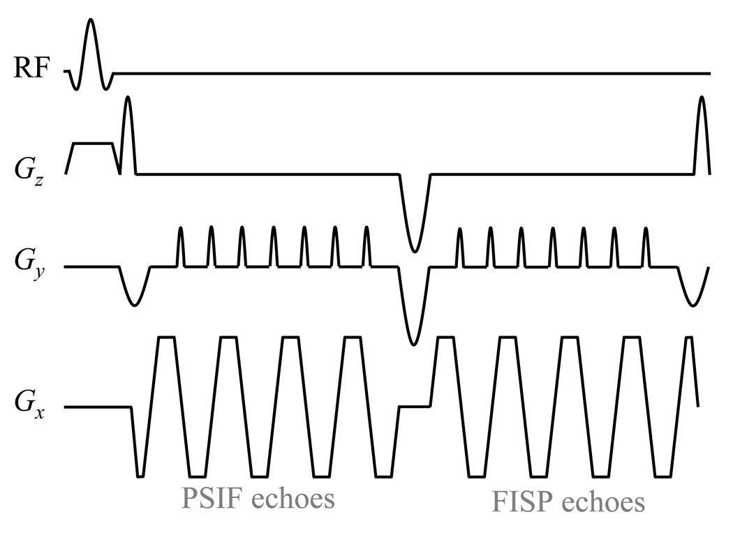
Fig. 4.
The gradient-echo EPI version of the pulse sequence design proposed here is depicted. The readout scheme from Fig. 2c is modified so that single-line readouts are replaced by EPI readouts instead. As in Fig. 2c, ‘superblips’ along the Gz(t) waveform allow PSIF echoes to be acquired first, followed by FISP echoes. Note that the first and last superblips are smaller than the middle one because they were merged with the negative rephaser and dephaser lobes associated with the slice-selective excitation pulse. Because the phantom imaged here was water-based and featured no fat signals, a regular RF pulse was employed, although, for in vivo imaging, it should be replaced with a spectral-spatial pulse.

