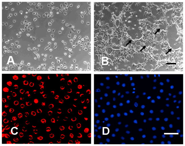
Figure 1. Characterization of pulmonary EC.
A. Phase contrast of pulmonary EC in culture isolated employing sequential anti-CD34 and anti-CD102 antibody selection. B. Characteristic tube-formation at confluence. C. Ac-LDL uptake assay. D. Cells in the same field as IC stained with DAPI for nuclear staining. Pulmonary EC display cobblestone morphology, form multicellular tubes (arrow) and express high levels of Ac-LDL confirming their endothelial phenotype. Size bar = 100μm

