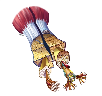
Figure 1:
Schematic view of the Achilles tendon. Left: Normal tendon showing a white glistening appearance, with tightly bundled type I collagen fibres, sparse cellularity and minimal vascularity. Right: Tendinopathic tendon showing a yellowish or greyish appearance, with underlying findings of tendinosis, which may include smaller and more disorganized fibrils, increased cellularity and vascularity, and increased proteoglycan (blue) and lipid (yellow) accumulation. Reproduced with permission from Brukner and Khan.1
Image courtesy of McGraw-Hill

