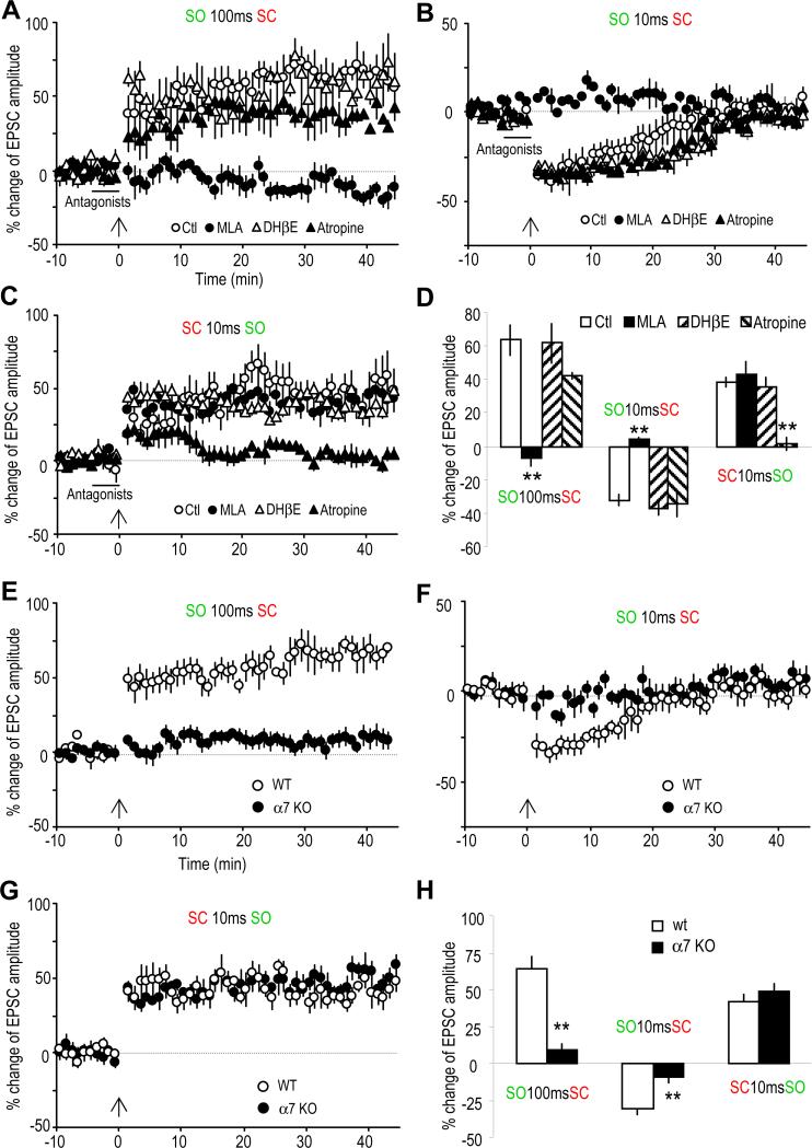
Fig. 2. Involvement of α7 nAChRs and mAChRs in mediating synaptic plasticity.
(A to C) % change of EPSC amplitude showing the effects of three cholinergic antagonists on the three types of plasticity. (A) The LTP induced by pairing the SO 100 ms before the SC was completely blocked by the α7 nAChR antagonist MLA (10 nM), but not by the non-α7 nAChR antagonist DHβE (1 αM) nor the mAChR antagonist atropine (5 μM). (B) The STD induced by pairing the SO 10 ms before the SC was also completely blocked by MLA, but not by DHβE nor atropine. (C) The LTP induced by pairing the SC 10 ms before the SO was blocked by atropine, but not by MLA nor DHβE. (D) Bar graph showing the effects of these three cholinergic antagonists on the three types of synaptic plasticity.
(E to G) % change of EPSC amplitude showing the difference in inducing synaptic plasticity in wild type (WT) and α7 nAChR knockout (KO) mice. (E) LTP was induced by pairing the SO 100 ms before the SC in WT, but not the α7 KO mice. (F) STD was induced by pairing the SO 100 ms before the SC in WT, but not α7 KO mice. (G) The LTP induced by pairing the SC 10 ms before the SO was not affected in the α7 KO mice. (H) Bar graph showing the difference between WT and α7 KO mice. **p<0.01, as compared with WT, student t-test, n=5-6.

