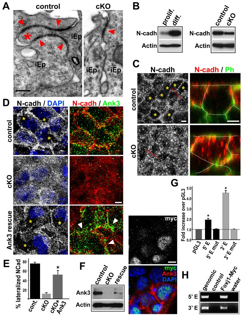
Figure 5. Foxj1 regulation of Ank3 during SVZ niche formation.
(A) TEM of P4 ventricular wall lateral junctions between immature ependymal progenitors (iEp). Note the lack of interdigitation (*) / extension of apical adherens junctions (arrowheads) in cKO lateral borders. (B) Western blot analyses comparing N-cadherin expression levels during in vitro differentiation of pRGPs. (C) N-cadherin (N-cadh) IHC staining of P4 ventricular wall wholemounts in X-Y and X-Z planes. Cell borders visualized by Phalloidin (Ph) (traced by dashed lines for clarity). Ph and DAPI were used as landmarks to ensure cytoplasmic scanning in X-Z planes. (D) IHC analyses of N-cadherin expression in Foxj1 cKO pRGPs infected with lentivirus expressing 190 kD Ank3. Note the clearance of N-cadherin protein (*) from the cytoplasm of Foxj1 cKO pRGPs expressing Ank3 (arrowheads). (E) % of differentiated pRGP in culture with lateralized N-cadherin staining. In Foxj1 cKO rescue experiments, we assessed N-cadherin status in cells expressing infected Ank3. * p < 0.05 Wilcoxon 2-sample test; error bar = stdev.; n = 5. (F) Western blot and IHC analyses of Ank3 expression in cKO pRGPs infected with lentivirus expressing Foxj1-Myc. (G) Transcriptional activity of 5′ and 3′ enhancer elements assayed by luciferase constructs in pRGP cultures. * p < 0.001 Wilcoxon 2-sample test; error bar: sem; n = 6. (H) PCR primer amplification of 5′ Enh and 3′ Enh genomic DNA fragments after chromatin immunoprecipitation with Myc antibody from differentiated pRGPs. Scale bar: (A) 0.5 μm; (C, D, F) 5 μm. See also Figure S6.

