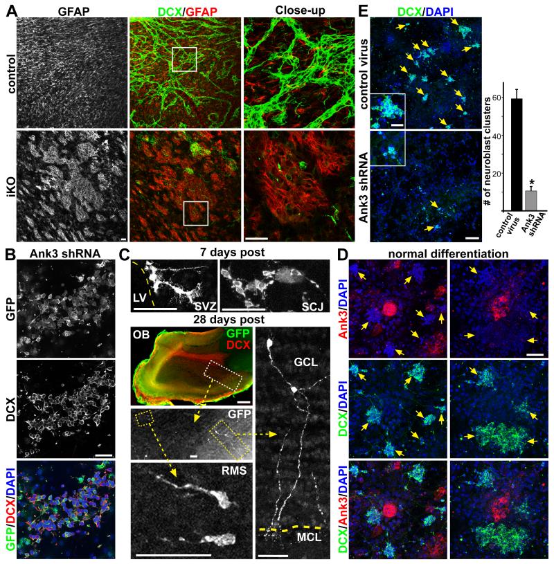
Figure 8. Ank3 expression in SVZ niche is required for neuroblast production.
(A) IHC staining of ventricular wholemounts from P28 control and iKO mice injected with tamoxifen at P14, showing abnormal GFAP+ patches in targeted areas. (B) SVZ NSC adherent culture from wild-type mice infected with lentivirus expressing Ank3 shRNA and GFP driven by ubiquitous EF1α promoter, showing abundant GFP+DCX+ neuroblasts 4 days after in vitro differentiation. (C) GFP staining of brain sections from mice transplanted with Ank3 shRNA-infected SVZ NSC culture, 7 and 28 days post transplantation. SCJ = striatal cortical junction; RMS = rostral migratory stream; GCL = granular cell layer; MCL = mitral cell layer. (D) IHC staining of wild-type pRGP niche cultures: 5 days after plating large numbers of DCX+ neuroblast clusters can be seen as well as Ank3+ niche progenitor clusters. (E) pRGP niche cultures infected with control versus Ank3 shRNA lentivirus, showing a dramatic reduction in the numbers of DCX+ neuroblast clusters (arrows), and quantified below (each cluster has greater than 5 DCX+ cells per DAPI staining, using same software acquisition as described in Figure S4B). * p < 0.01 Wilcoxon 2-sample test; error bar = stdev.; n = 5. Scale bar: (A) 50 μm; (B) 25 μm; (C) OB 500 μm, all others 50 μm; (D) 50 μm; (E) 100 μm / close-up 25μm.

