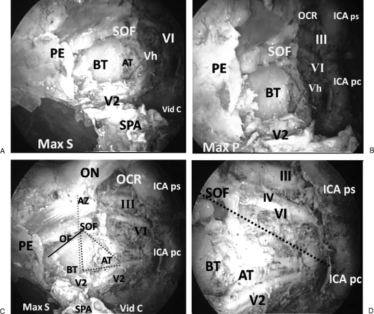
Figure 2.
(A) Endoscopic view of right maxillary nerve (V2) after drilling its canal superolateral to cut sphenopalatine artery (SPA). The superior orbital fissure (SOF) and orbital apex become more evident. Adipose tissue (AT) is partially obscuring a huge vein (Vh), superior ophthalmic vein (SOV), above V2. The maxillary sinus (Max S) posterior wall and remnant of posterior ethmoid sinus (PE) appear intact showing that the pterygopalatine fossa is not violated. A bone trajectory (BT) separates V2 canal from SOF at the cavernous sinus apex. Vidian canal (Vid C) protuberance appears in the floor of sphenoid sinus. Abducent nerve (VI) appears in anterior part of the medial wall of cavernous sinus. (B) Endoscopic view of V2 in its canal in relation to the medial wall of parasellar region. After removal of adipose tissue above V2, a huge vein (Vh), namely the SOV, appears originating from SOF and passing parallel to V2. A small tributary accompanying V2 in its canal is joining SOV. The occulomotor (III) and abducent (VI) nerves emerge laterally to the parasellar internal carotid artery (ICA ps) and pass anteriorly toward the SOF. Posteriorly, the cavernous internal carotid artery (ICA) extends downward from opticocarotid recess (OCR) as parasellar (ICA ps) and paraclival (ICA pc) segments. (C) Endoscopic view showing the four sides of the approach. V2 inferiorly, optic nerve (ON) in its dural sheath superiorly, and cavernous ICA posteriorly, which extends from opticocarotid recess (OCR) to V2 decussation with ICA. The cavernous ICA is formed by a C shaped parasellar segment (ICA ps) and a vertical paraclival segment (ICA pc). The anterior side is shown with a vertical dashed line passing through annulus of Zinn (AZ), SOF, bone trajectory between SOF and V2, and its canal. After removal of bone covering the medial wall of parasellar region and dura forming the medial of cavernous sinus, the abducent (VI) and occulomotor (III) nerves can be seen in the cavernous sinus apex anteriorly. Posteriorly, these nerves lie lateral to the ICA ps. AT above V2 obscures ophthalmic nerve (V1). The Vid C appears in the floor of sphenoid sinus and passes anterolateral to SPA and foramen, where it is very close to V2. The posterior wall of Max ) appears anterior to V2 canal. The cavernous sinus apex is exposed by drilling a bony dashed triangle limited by V2 and its canal inferiorly, V1 posterolaterally, and BT between SOF and V2 canal anteriorly. This BT becomes narrower anteriorly due to downward slope of orbital floor (OF) (continuous line) anteriorly, favoring a mediolateral approach to SOF. (D) Endoscopic view showing nerves in the medial wall of right cavernous sinus. The medial wall of cavernous sinus is divided by a dashed line into motor and sensory triangles. In the motor triangle (top) the abducent nerve (VI) is the most superficial, and the occulomotor (III) is superior and parallel to VI and at a deeper level. The trochlear nerve is barely seen deep and parallel to VI and inferior to III. The sensory triangle (bottom) contains V2 and AT, which obscures a huge SOV and ophthalmic nerve (V1) at a deeper lateral level.

