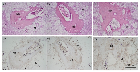
Fig. 4.
HE staining (a–c) and IHC staining (d–f) of serial sections of Sample A after 30-d implantation
(a, d) Primary antibody of anti-mouse BMP-2; (b, e) Primary antibody of anti-mouse collagen type I; (c, f) Primary antibody of anti-mouse osteopontin. M: decalcified biomaterials; NB: induced bone. Brown colored area: positive region

