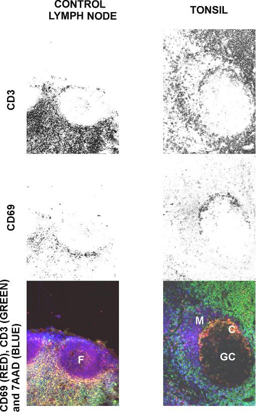
Figure 4.

Comparison of the distribution of activated and memory T cells in frozen sections from human control lymph node and tonsil. Sections were stained with 7 AAD, anti-CD69-PE and with anti-CD3-FITC monoclonal antibodies. F: primary follicle; GC: germinal centre; M: mantle of B cells; C: centrogerminal layer of T cells.
